
Cardiac Imaging with AI: Uses, Benefits and What Is Next
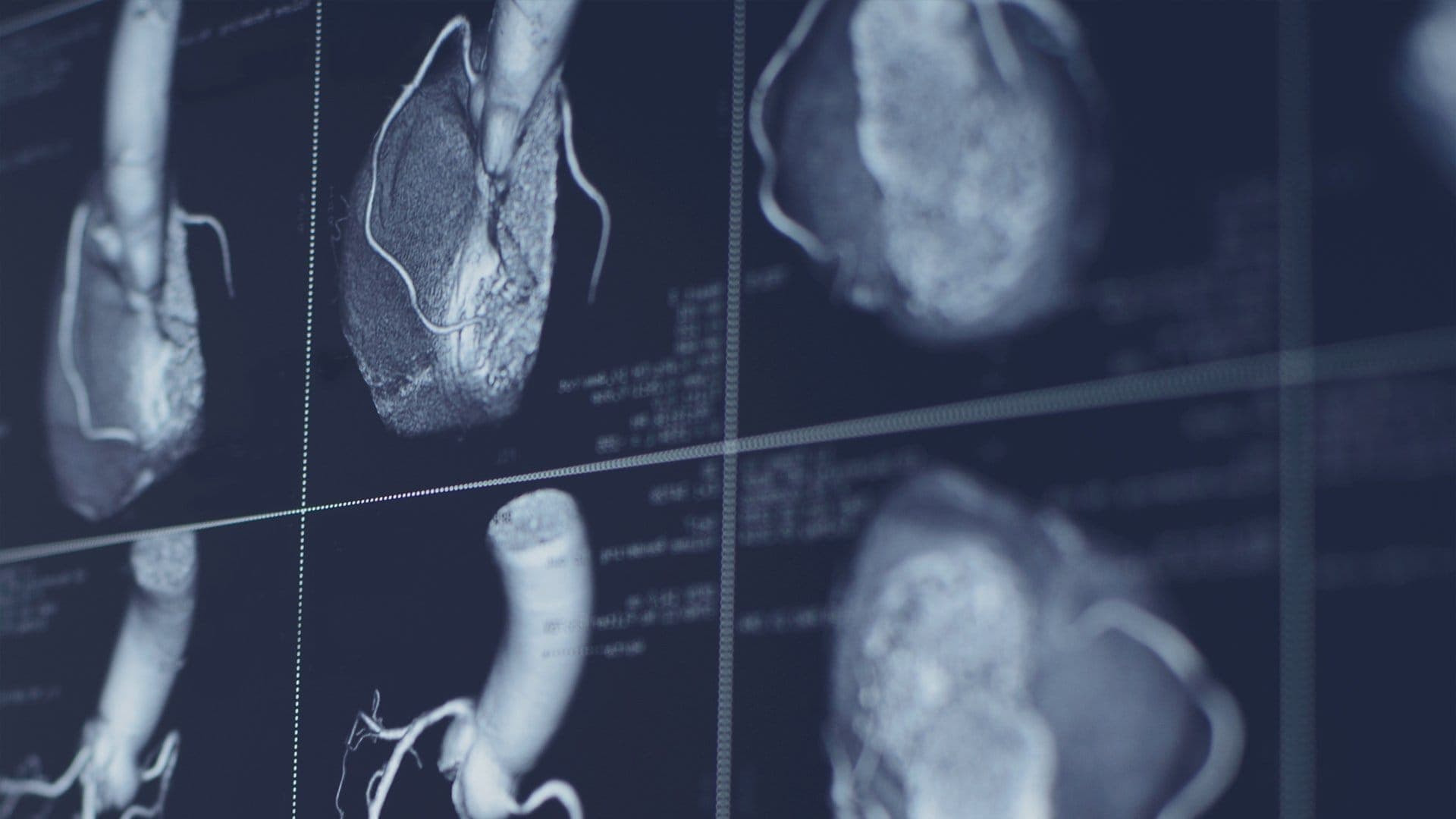
Cardiac Imaging with AI: From Film to Intelligent Diagnostics
AI in cardiac imaging is used to acquire cleaner images faster, measure structures such as ejection fraction automatically, flag urgent findings for priority review, and draft structured reports from study data. It works inside the PACS and reporting workflow as decision support, giving cardiologists a consistent first read while the final interpretation stays with the clinician.
The Evolution of Cardiac Imaging: Film, Digital, Intelligent
Cardiac imaging began with film and fluoroscopy. A study lived on physical media, it could sit in one room at a time, and comparing a new scan with a prior one meant retrieving folders and hoping nothing was missing. Echocardiography added real-time views of the beating heart, but early studies were still captured, stored, and shared through slow, manual processes.
The digital era changed the foundation. DICOM standardized how imaging devices store and exchange studies, and PACS replaced film libraries with searchable digital archives. Cardiac CT and cardiac MRI matured into routine tools, producing detailed views of coronary arteries, valves, and myocardial tissue. Cardiologists could pull up current and prior studies side by side from any workstation.
The current era adds intelligence to that digital base. Imaging volume keeps growing, studies keep getting larger, and cardiology teams face the same reporting deadlines with more data per patient. AI addresses that gap. It does not replace the imaging modalities cardiologists rely on. It makes each stage of the workflow, from acquisition to final report, faster and more consistent.
How AI Is Used in Cardiac Imaging Today
Image Acquisition and Reconstruction
AI now works at the scanner and the ultrasound machine, before a human ever opens the study. Reconstruction algorithms reduce noise and motion artifacts, producing cleaner images from lower-dose cardiac CT scans and shorter cardiac MRI acquisitions. On echocardiography systems, AI guides probe positioning and checks image quality in real time, so a usable study is captured the first time instead of calling the patient back.
For the reading cardiologist, the practical effect is simple: fewer nondiagnostic studies, less repeat imaging, and cleaner source images for interpretation.
Automated Measurements
Measurement is where AI saves the most routine time. Algorithms outline cardiac chambers, calculate ejection fraction, measure wall thickness and volumes, and quantify valve areas in seconds. In echocardiography, automated ejection fraction and strain analysis replace minutes of manual tracing with a consistent starting point the cardiologist can verify and adjust.
Consistency matters as much as speed. Two readers measuring the same study by hand can produce noticeably different numbers. An automated first measurement gives every reader the same baseline, which makes serial comparisons, such as tracking ejection fraction across a course of treatment, far more reliable.
Plaque and Calcium Analysis
AI tools analyze coronary CT angiography to identify and characterize plaque, estimate stenosis severity, and support coronary calcium scoring. Calcified and noncalcified plaque can be separated and quantified across the coronary tree, turning a long manual review into a structured summary the cardiologist confirms.
This matters because plaque burden drives treatment decisions. A consistent, quantified read of coronary calcium and plaque helps cardiologists stratify risk and document findings in a form that referring physicians can act on quickly.
Triage and Prioritization
Not every study carries the same urgency. AI triage tools scan incoming studies and flag findings that need fast attention, such as a severely reduced ejection fraction or signs of acute pathology, and move them to the top of the worklist. Routine studies wait their turn. Critical ones do not sit unopened in a queue behind a backlog of normal results.
Connected to the PACS worklist, triage turns AI from an analysis tool into a patient safety tool. The sickest patients get read first, and turnaround time improves where it matters most.
Structured Reporting
Findings only help if they reach the chart in a usable form. AI reporting tools pull measurements and detected findings into structured report templates, so the cardiologist edits and signs a drafted report instead of dictating every value from scratch. Structured data also flows cleanly into the EHR through HL7 and FHIR interfaces, where it can be searched, trended, and shared instead of being buried in free text.
Portability: Cardiac Imaging at the Point of Care
The second major shift is where imaging happens. Handheld and point-of-care ultrasound devices now put a capable cardiac assessment at the bedside, in the emergency department, in a rural clinic, or during a home visit. A clinician can check for heart failure, pericardial fluid, or gross wall motion problems in minutes, then decide whether the patient needs a full study.
Portable devices only reach their full value when the studies they capture join the same record as everything else. Images from a handheld device need to land in the PACS, attach to the right patient, and appear in the same worklist as cart-based echocardiography. A cloud PACS makes that possible: studies upload from wherever they were acquired, and a cardiologist across town or across the state can review them remotely, with full image quality, from a browser.
For underserved areas, this closes a real gap. The scan happens locally, the expertise is applied remotely, and the patient avoids a long trip for a first assessment.
AI strengthens portable imaging further. Handheld devices increasingly include on-device guidance that tells the operator how to angle the probe, scores the view quality in real time, and captures diagnostic-grade loops with less formal training. That lowers the skill barrier at the point of care while the reading still happens in the same PACS workflow. A nurse or general physician in a small clinic can acquire a usable cardiac study, and a cardiologist reviews it remotely with the same tools used for in-lab echocardiography. Portability stops being a compromise on quality and becomes a genuine extension of the imaging department.
Ultra-High Resolution Imaging: Seeing More, Earlier
Resolution is the third shift. Photon-counting CT, the newest generation of cardiac CT, counts individual X-ray photons and converts them directly into image data. The result is sharper images with better contrast at lower radiation dose, revealing small stents, fine calcification detail, and subtle plaque features that conventional detectors blur. High-resolution cardiac MRI has advanced in parallel, with faster sequences and finer spatial detail for tissue characterization, scar imaging, and myocarditis assessment.
What this changes clinically is timing. Subtle disease becomes visible earlier: early atherosclerotic plaque before it produces symptoms, small areas of myocardial scar, early cardiomyopathy changes. Earlier detection means earlier intervention, when treatment has the most effect.
Echocardiography has moved in the same direction. Three-dimensional echo now renders valves and chambers as volumetric models instead of flat slices, and high-frame-rate imaging captures fast-moving structures such as valve leaflets with far less blur. Combined with automated quantification, these advances let cardiologists plan interventions, from valve repair to device placement, with measurements taken directly from the study rather than estimated from a single plane. Resolution gains are no longer confined to the CT and MRI suites. They are changing what a routine echo can answer.
The trade-off is data. Ultra-high resolution studies are large, and they multiply quickly. Storage, fast retrieval, and a viewing workflow that does not stall on multi-gigabyte studies become part of the imaging decision, which is why resolution upgrades and cloud PACS upgrades increasingly happen together.
Benefits of AI in Cardiac Imaging
The benefits cluster into three areas. Accuracy support comes first: AI applies the same measurement method to every study, catches patterns a tired reader might pass over late in a long list, and gives cardiologists a second set of eyes on every case. Workflow speed follows: automated measurements, drafted reports, and triage shorten the path from acquisition to signed report, which relieves the reporting backlog most cardiology departments carry. Finally, access improves: portable acquisition, cloud review, and AI guidance during scanning let smaller practices offer cardiac imaging services that once required a large hospital imaging department.
Limitations and Risks: Where AI Still Needs Oversight
AI in cardiac imaging has real limits, and practices should plan around them. Algorithms perform best on the kinds of cases they were trained and validated on. Unusual anatomy, rare conditions, poor-quality source images, and patient populations underrepresented in training data can all produce unreliable output. A confident automated measurement can still be wrong.
Validation and oversight are therefore part of the workflow, not an optional extra. Practices should confirm that any AI tool they use has appropriate regulatory clearance for its stated purpose, test it on their own case mix before relying on it, and keep the cardiologist as the final reader on every study. AI output also has to move through HIPAA-compliant systems with the same access controls and audit trails as the rest of the imaging record. The safe model is consistent: AI prepares, measures, and prioritizes, and the clinician decides.
What This Means for Cardiology Practices
For a cardiology practice, these three shifts, AI, portability, and resolution, land as infrastructure questions. Can the PACS store and retrieve ultra-high resolution studies without slowing readers down? Do portable studies from every location flow into one worklist? Do AI measurements and reports connect to the EHR without manual re-entry? Does the system meet HIPAA requirements for every image, wherever it was captured?
Practices that answer yes get compounding returns: faster reports, consistent measurements, and imaging services that reach patients where they are. Practices running aging on-premise systems usually feel the ceiling first in storage limits, slow remote access, and disconnected reporting.
How SARC MedIQ Supports AI-Driven Cardiac Imaging
SARC MedIQ brings these pieces together in one cloud platform. PACSflow, its cardiology PACS (CPACS), stores and serves high-resolution cardiac studies without local servers, so echocardiography, cardiac CT, and cardiac MRI studies are available from any location with the same worklist. AIReports applies AI assistance to findings and structured reporting, turning measurements and detected findings into drafted reports cardiologists review and sign. AIDictate adds voice-driven reporting for clinicians who prefer to dictate, converting speech into structured report text.
Because the platform is cloud-based and DICOM-native, portable and point-of-care studies upload into the same archive as in-lab studies, and reports move to the EHR through standard interfaces. The result is one workflow for acquisition, AI-assisted reading, reporting, and secure sharing, whether the study started in a cath lab, a clinic, or at the bedside.
Conclusion
Cardiac imaging has moved from film, to digital archives, to intelligent workflows, and each step widened what cardiology teams can see and how fast they can act on it. AI measurement and triage, portable point-of-care devices, and ultra-high resolution modalities like photon-counting CT are not separate trends. They work best as one connected system, supported by a cloud PACS that keeps every study, measurement, and report in a single workflow.
If your practice is weighing how to bring AI into its cardiac imaging workflow, book a 30-minute demo to see how SARC MedIQ handles acquisition, AI-assisted reporting, and remote review in one platform.
Frequently asked questions
AI is used in cardiac imaging to reconstruct cleaner images, automate measurements such as ejection fraction and chamber volumes, analyze plaque and coronary calcium, flag urgent studies for priority reading, and draft structured reports. It sits inside the acquisition device, the PACS, and the reporting workflow as decision support. The cardiologist reviews, corrects, and signs off on every result, so AI speeds up the routine work without replacing clinical judgment.
No. AI supports cardiologists rather than replacing them. It handles repetitive tasks like tracing chambers, calculating ejection fraction, and sorting worklists by urgency, and it applies a consistent method to every study. Interpretation, diagnosis, and treatment decisions remain with the cardiologist, who also catches the unusual cases and poor-quality images where automated output can be unreliable. Used this way, AI raises consistency and speed while clinical responsibility stays exactly where it belongs.
Photon-counting CT is a newer form of cardiac CT that counts individual X-ray photons instead of averaging their energy. It produces sharper images with better contrast at a lower radiation dose than conventional CT. For cardiology, that means clearer views of coronary arteries, small stents, calcifications, and plaque features that older scanners blur. Earlier, more detailed detection of coronary disease supports earlier treatment decisions. The trade-off is much larger study files, which require strong PACS storage and retrieval.
Point-of-care ultrasound lets clinicians perform focused cardiac assessments at the bedside, in emergency departments, and in remote clinics using handheld or portable devices. It answers immediate questions, such as whether heart failure or fluid around the heart is present, and helps decide who needs a full echocardiography study. It fits the wider workflow when the images upload into the same cloud PACS and patient record as lab-based studies, so findings are documented, reviewable remotely, and available for comparison later.
Start with validation: confirm the tool is cleared for its stated use and test it on your own case mix before relying on its output. Check integration next, so measurements and reports flow into your PACS and EHR through DICOM, HL7, or FHIR instead of creating a separate silo. Review the security model for HIPAA compliance, access controls, and audit trails. Finally, confirm the workflow keeps a cardiologist as the final reader on every study.

Asaad Hakeem, Ph.D.
Asaad Hakeem, Ph.D., is the Chief Executive Officer of SARC MedIQ and a leader in applying artificial intelligence to healthcare imaging and clinical workflows. With a strong background in machine learning, computer vision, and AI research, he has focused his career on solving real-world challenges in medical data accessibility and diagnostic efficiency. Asaad Hakeem is also an active member of the Forbes Business Council and was selected to lead its Healthcare Group, reflecting his influence in advancing healthcare innovation. Under his leadership, SARC MedIQ has developed a cloud-based imaging platform that helps healthcare providers streamline reporting, improve accuracy, and enhance patient care. His work emphasizes practical, scalable solutions that integrate seamlessly into clinical environments. He continues to contribute thought leadership on healthcare transformation, AI adoption, and value-based care models.

Never miss an insight that matters
Subscribe to get the latest articles, case studies, and product updates delivered straight to your inbox.
Ready to modernize your practice?
Discover how MedIQ and optional modules can streamline your workflow, improve efficiency and modernize your practice.
Book 30-min demo