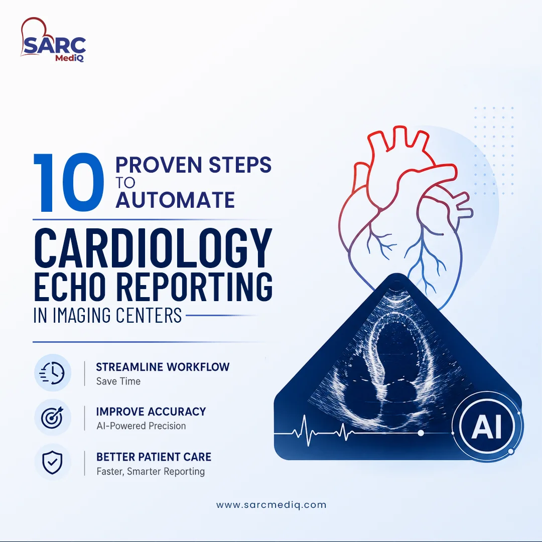
10 Proven Steps to automate Cardiology Echo Reporting in Imaging Centers
Automation is rapidly reshaping how imaging centers deliver cardiology services. Echo and stress test reporting has long been plagued by manual measurements, inconsistent templates, and data silos. These tests can now be accelerated using AI-powered, structured reporting systems. For imaging centers, the question is not whether automation will happen, but how to implement it efficiently, safely, and cost-effectively.
This guide outlines ten proven steps to automate cardiology echo reporting, from mapping workflows to scaling across the enterprise, helping centers achieve faster turnaround, fewer errors, and greater clinical consistency.
SARC MedIQ – purpose built for cardiovascular and vein practices
SARC MedIQ leads to the transformation of cardiac imaging in outpatient and independent practice settings. The platform provides a clinically focused, cloud-native suite that automates echo and stress test reporting while remaining simple to deploy and vendor-neutral.
Unlike enterprise-heavy systems, SARC MedIQ is purpose-built for practices seeking measurable time savings, consistent quality, and flexible per-exam pricing.
The platform connects seamlessly with existing ultrasound systems and PACS, capturing data automatically, generating structured echo reports, and enabling secure collaboration through ShareSecure.
With deep specialization in cardiovascular, vein, and stroke imaging, SARC MedIQ shortens report turnaround times, reduces transcription overhead, and maintains compliance with guideline-based templates—all without disrupting existing workflows. The result is an AI-native reporting workflow that gives clinicians their time back while enhancing diagnostic quality.
Following are 10 steps any practice must follow to automate Cardiology Echo Reporting in Imaging Centers.
1. Map current workflow and set performance targets
Before automation begins, understanding current workflows is essential.
Start by quantifying average study volumes, report turnaround times, and backlog metrics. Map each integration point—PACS, EMR, modality worklists—and identify where delays or data re-entry occur.
Once pain points are visible, define measurable improvement goals such as cutting report turnaround by 40% or reducing manual measurement entry by half. These targets will guide the automation strategy and provide tangible outcomes to demonstrate operational and clinical improvements.
2. Define automation scope and clinical requirements
Echo reporting automation can range from partial assistance to complete AI-driven generation.
Three main approaches include:
- Measurement-assisted reporting: Automatically imports DICOM SR data into structured templates, reducing manual entry.
- AI-guided drafts: Generates preliminary structured impressions for cardiologist validation.
- End-to-end automation: Produces comprehensive reports, including disease detection, with clinician oversight.
Structured reporting relies on standardized templates with defined parameters—such as chamber measurements, valve grades, and LVEF—to promote consistency and accuracy.
Choosing the right automation depth depends on clinical complexity, case volume, and staff readiness. Platforms like SARC MedIQ support all three approaches to match your desired automation level and clinical confidence.

3. Prioritize vendor-agnostic device compatibility
A vendor-agnostic solution protects existing hardware investments and ensures scalability. Many centers operate mixed ultrasound fleets; therefore, automation software should support all major vendors while adhering to DICOM SR and HL7 standards.
| Evaluation Criterion | Ideal Solution Characteristic |
| Vendor neutrality | Supports GE, Philips, Siemens, Canon, Mindray |
| Device support | Any DICOM SR-compliant ultrasound |
| PACS/EMR integration | HL7 bi-directional, cloud or on-prem compliant |
Browser-accessible, vendor-neutral tools such as SARC MedIQ make it possible to implement automation without replacing legacy systems or engaging in costly custom interfaces.
4. Assess regulatory clearance and clinical evidence
When integrating AI tools into diagnostic workflows, regulatory clearance is non-negotiable. Confirm FDA 510(k) or CE marking for any automation software, and review published evidence validating safety and accuracy.
AUC (Area Under the Curve) measures diagnostic performance i.e. values closer to 1.0 indicate high reliability. Systems such as PanEcho have demonstrated AUC values approaching 0.98–1.00 for disease recognition.
Platforms like US2.AI, a SARC MedIQ partner, report time reductions from approximately 35–40 minutes to 10 minutes per study, offering strong evidence of both efficiency and accuracy improvements.
SARC MedIQ complements these standards by maintaining transparent quality metrics and clinician oversight throughout every AI-assisted report.
5. Plan integrations with EMR and Modality worklists Early
Integration defines the success or failure of automation projects. Begin with a clear interface plan covering EMR and modality worklists. Typical interface setups may cost $2,000–$10,000 per modality depending on vendor complexity.
A best-practice integration sequence includes:
- Gather integration requirements with clinical and IT teams.
- Validate device and PACS compatibility.
- Conduct a limited pilot to confirm data flow.
- Complete phased rollout, with early monitoring and clinician validation.
Planning integration early avoids cost overruns and operational disruption. Cloud-native platforms like SARC MedIQ streamline this process with standardized HL7 and DICOM connectors that eliminate much of the typical interface friction.
6. Run a phased pilot with clinician involvement
Pilot studies build clinician confidence while surfacing usability issues. Begin with measurement auto-population or AI-generated drafts and retain cardiologist sign-off for each report.
Collect data on turnaround times, error frequency, and report edits. Controlled trials—such as NCT07229300—have shown that AI-preliminary echoes can achieve diagnostic concordance levels comparable to human preliminaries, reinforcing the value of structured pilot testing before full deployment. S
ARC MedIQ’s implementation model routinely includes clinician-led pilots to validate measurable gains before scaling.
7. Configure templates and align to clinical guidelines
Standardized templates aligned with cardiology society guidelines maintain diagnostic consistency across users. Design templates that automatically populate key fields like LVEF, valve morphology, and chamber dimensions.
Essential parameters for guideline-aligned templates include:
- Patient demographics and clinical indications
- LV/LA/RV size and function
- LVEF or strain analysis
- Valvular assessment and Doppler findings
- Summary impressions and recommendations
Preloaded structured templates reduce variation and enhance auditability. SARC MedIQ includes built-in templates mapped to ASE and IAC-accredited standards for faster, compliant echo documentation.
8. Implement Quality Assurance feedback loops
Automation is not a “set and forget” process. Continuous quality assurance ensures accuracy and clinical safety. Routinely compare automated outputs against physician-edited final reports.
Periodic audits should track:
- Discrepancy rates
- Frequency of clinical edits
- Misinterpretation or data omissions
Recent studies show that large language model–generated echo reports are clinically acceptable in over 85% of cases, with minimal parameter discrepancies, underscoring the need for continuous supervision and AI-threshold tuning.
SARC MedIQ incorporates audit-ready tracking and feedback dashboards to simplify this process for compliance reviewers and medical directors.
9. Train staff and establish governance processes
Effective automation requires multidisciplinary ownership. Provide hands-on training for sonographers, cardiologists, and system administrators before go-live. Establish clear escalation protocols for ambiguous findings and define final report accountability.
Governance should also cover:
- Version control for templates and AI logic
- Review cadence for system updates
- Documentation of all protocol or model changes
These safeguards preserve medico-legal clarity and clinician trust. SARC MedIQ’s onboarding framework standardizes these governance elements to ensure both clinical safety and adoption of success.
10. Scale automation with monitoring and cost controls
Once proven in one site, gradual scaling ensures sustained ROI. Track post-deployment metrics—reporting turnaround, error rates, user satisfaction, and storage costs.
Cloud reporting platforms such as SARC MedIQ or Tempo can cost as little as $270–$345 per month for small centers, compared with $800–$1,500 per provider on some enterprise systems. Regular cost reviews, coupled with optimization of cloud subscriptions and maintenance, sustain profitability as automation expands across multiple locations.
SARC MedIQ also enables per-exam pricing—keeping advanced AI automation accessible without heavy capital investment.
Frequently Asked Questions on Echo Reporting
What are the key steps to successfully implement AI automation in echo reporting?
Map workflows, define automation depth, select vendor-agnostic tools such as SARC MedIQ, ensure regulatory compliance, plan integrations early, pilot with clinicians, align templates to guidelines, implement QA, train teams, and scale with cost monitoring.
Which tools or AI features help automate echo measurements like ejection fraction and strain?
SARC MedIQ automatically calculates ejection fraction, strain, and chamber volumes from echo images to standardize reporting and minimize manual input.
How can imaging centers ensure regulatory compliance with AI-powered echo reporting?
Select platforms with regulatory clearance (FDA, CE), maintain complete documentation, and ensure clinician sign-off for every AI-assisted report. SARC MedIQ provides compliance-ready templates and audit logs aligned with accreditation standards.
What hardware and software infrastructure are needed to support echo reporting automation?
A cloud-connected PACS and standard DICOM/HL7 connectivity are typically sufficient; additional hardware is not required beyond ultrasound machines and secure workstations.
What time savings and clinical benefits can imaging centers expect from automation?
AI-driven echo reporting can cut report completion time by up to 70%, reduce manual errors, and give clinicians more time to focus on patient care rather than administrative work.