How AI Cuts Echo and Stress Test Reporting Time by Half
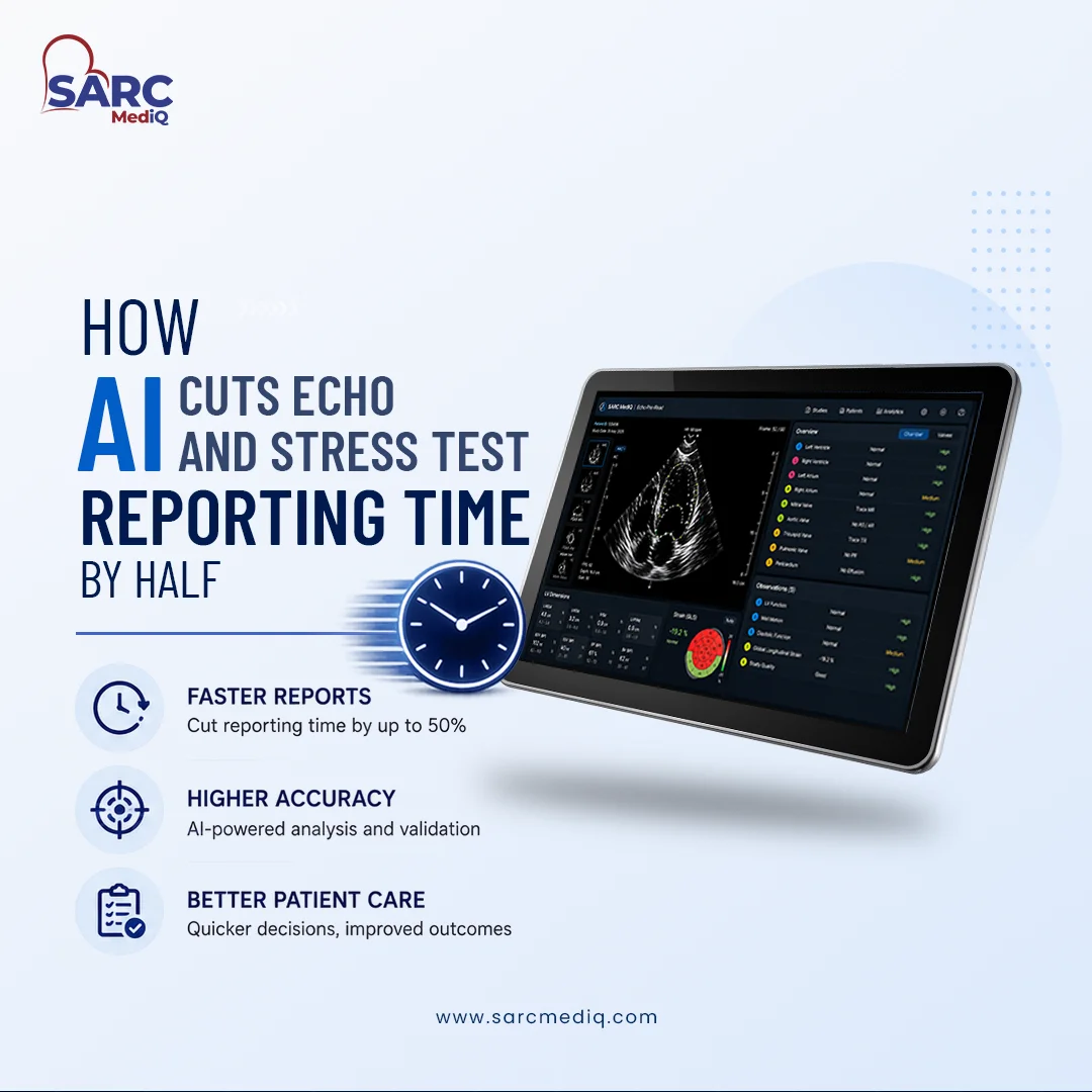
Artificial intelligence is reshaping cardiovascular imaging by tackling one of clinicians’ biggest bottlenecks — report generation. Traditionally, echocardiogram and stress test reporting require extensive manual measurements and narrative entry. With AI-powered automation, those tasks are now completed in minutes, reducing average echo and stress test reporting time by as much as 50%–70%. For outpatient and specialty practices, the result is not just faster turnaround but a more standardized, reliable, and less stressful workflow. Platforms like SARC MedIQ, a cloud-native, AI-driven imaging workflow solution, exemplify how AI can augment human expertise while maintaining compliance, interoperability, and diagnostic quality.
Overview of AI Impact on Cardiovascular Imaging Reporting
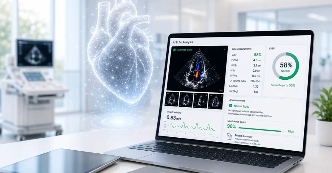
Artificial intelligence in cardiovascular imaging uses machine learning algorithms to interpret ultrasound and stress test data, automatically measure cardiac structures, and generate structured reports. By automating core measurements such as ejection fraction, wall thickness, and chamber volumes, AI eliminates repetitive manual steps that slow down cardiologists and sonographers.
Peer-reviewed evaluations show that AI-powered reporting systems can cut echocardiography reporting time by up to 70%, translating to a reduction of more than 500 seconds per study. These gains compound across daily case volumes, allowing mid-sized practices to handle more patients without increasing staffing or compromising quality. As a cloud-native platform, SARC MedIQ applies AI-native workflow automation tuned to the needs of independent clinics—delivering measurable improvements in cardiology reporting efficiency and echo turnaround times.
Key AI Capabilities Accelerating Echo and Stress Test Reporting
Three categories of AI capability drive most of the efficiency gains:
- Automated view classification and segmentation: Sophisticated algorithms can recognize echocardiographic views in milliseconds and accurately identify anatomical boundaries. Studies show accuracy approaching 97.8% compared to clinical experts.
- Automated quantitative measurement extraction: Parameters such as ejection fraction (the percentage of blood ejected from the heart during each contraction), strain, and ventricular volumes are computed automatically, producing reproducible results without manual tracing.
- Auto-generated report drafts: Once images and measurements are processed, the system constructs a prepopulated, structured draft report ready for clinician review and final edits.
Workflow task distribution:
| Task | AI Action | Clinician Oversight |
| Image view recognition | Fully automated | Quality confirmation |
| Measurement extraction | Automated for standard views | Review outliers or poor-quality images |
| Report generation | Prepopulated structured report | Interpretation and sign-off |
Common Imaging Software Used in Cardiovascular Clinics
Cardiovascular clinics rely on integrated imaging systems combining PACS (picture archiving and communication systems), vendor-neutral archives, and reporting software. Cloud-based solutions like SARC MedIQ, along with established options such as Sectra Cardiology and Infinitt, serve as the foundation for echo, stress test, and cath lab reporting.
Clinics increasingly prioritize:
- AI-enabled auto-measurements and quality control
- Structured templates aligned with regulatory requirements
- Seamless integration with EHR and modality equipment
| Modality | Traditional Reporting | AI-Enabled Reporting |
| Echocardiogram | Manual measurement and dictation | Auto-calculated metrics, prefilled draft report |
| Stress Test | Manual results entry | Real-time data capture, automated summary |
| Cardiac Cath | Manual report creation | Emerging AI modules for case structure guidance |
This hybrid model supports vendor-neutral adoption of Cloud PACS and AI cardiology reporting tools like SARC MedIQ, with minimal workflow disruption and without costly upgrades.
AI Tools That Reduce Echocardiogram and Stress Test Reporting Time
SARC MedIQ integrates proprietary AI modules that directly reduce the time required for both measurements and reporting:
- Automated measurement extraction: Completes common measurements such as LV dimensions and ejection fraction within ~159 seconds, compared to 325 seconds manually.
- AI-assisted report generation: Builds structured reports in around 71 seconds, versus 429 seconds in manual workflows.
- Automated quality control: Identifies suboptimal image acquisition before report finalization, improving consistency and reducing callbacks.
In comparative trials, these capabilities result in over 70% faster overall workflow completion. Other validated AI platforms show similar performance, with echo view recognition times as fast as 21 milliseconds per frame. Time reductions occur most sharply in measurement and draft report creation, where AI reliably standardizes repetitive processes.
Clinical and Operational Benefits of AI-Driven Reporting
Beyond speed, AI reporting tools deliver measurable improvements for staff and patients alike. Clinics implementing AI workflows typically experience:
- Higher case throughput: More studies completed per day without extra staff.
- Reduced sonographer fatigue: Repetitive measurement and manual entry replaced with streamlined verification.
- Improved consistency: AI-generated measurements show less operator variability and higher reproducibility.
- Enhanced diagnostic sensitivity: In stress echocardiography, AI support improves coronary artery disease detection rates by more than 10%.
- Elevated confidence: AI-guided measurements reinforce reliability for less experienced readers.
Together, these benefits improve diagnostic precision and help clinicians refocus time on patient care. Practices using SARC MedIQ often cite the added relief of predictable, high-quality reporting supported by transparent AI oversight.
Challenges and Considerations When Implementing AI in Cardiovascular Imaging
AI effectiveness depends heavily on data quality and responsible implementation. Poor-quality or incomplete image studies still require manual correction, reducing automation benefits. Clinicians also remain responsible for interpretation—AI does not replace human expertise.
Challenges include:
- Maintaining clinician trust and avoiding workflow disruption during rollout
- Ensuring compliance with HIPAA and FDA regulations
- Addressing data privacy and security frameworks
- Providing ongoing quality assurance through human validation
A human-in-the-loop approach, where clinicians confirm or edit AI-generated findings, remains the standard for safety and accountability. SARC MedIQ embeds these safeguards within its platform, integrating review protocols that maintain both speed and clinical integrity.
Best Practices for Integrating AI into Echo and Stress Test Workflows
Successful AI adoption follows a deliberate and incremental path. Clinics should:
- Assess workflow gaps and choose validated AI modules suited to their existing infrastructure.
- Implement in phases, starting with measurement automation before expanding to full report automation.
- Train staff regularly, emphasizing consistent image acquisition to maximize AI accuracy.
- Set human review protocols for clinical oversight and error detection.
- Monitor outcomes and refine, using performance metrics to continuously validate and improve AI usage.
Following these steps minimizes risk, builds staff confidence, and ensures sustained efficiency gains. SARC MedIQ supports this process through clinician-centered onboarding and live implementation support that accelerates adoption with minimal disruption.
Frequently Asked Questions
What imaging software do cardiovascular clinics use for echo, stress test, and cardiac cath reporting?
Clinics typically use cloud-based PACS and structured reporting platforms such as SARC MedIQ, which integrate seamlessly with ultrasound and cath lab systems to streamline documentation.
Which AI tools help cardiovascular clinics reduce echocardiogram and stress test reporting time?
AI features like automated measurement extraction, view classification, and instant draft report generation—core to SARC MedIQ—can reduce reporting time by up to 70%.
How accurate are AI-driven measurements and classification in echocardiography?
Validated models deliver up to 97.8% accuracy in view classification and produce quantitative measurements consistent with clinician performance.
Does AI eliminate the need for human review in cardiac imaging reports?
No. Clinicians always review and confirm AI-generated reports to ensure diagnostic accuracy, a process fully supported by SARC MedIQ’s human-in-the-loop design.
Does image quality affect AI reporting time savings?
Yes. High-quality image acquisition maximizes AI efficiency, while poor studies may require additional manual verification. SARC MedIQ includes automated quality checks to help identify and resolve these cases early.